Dr. Curtis Cripe: Common Tools for Brain Mapping
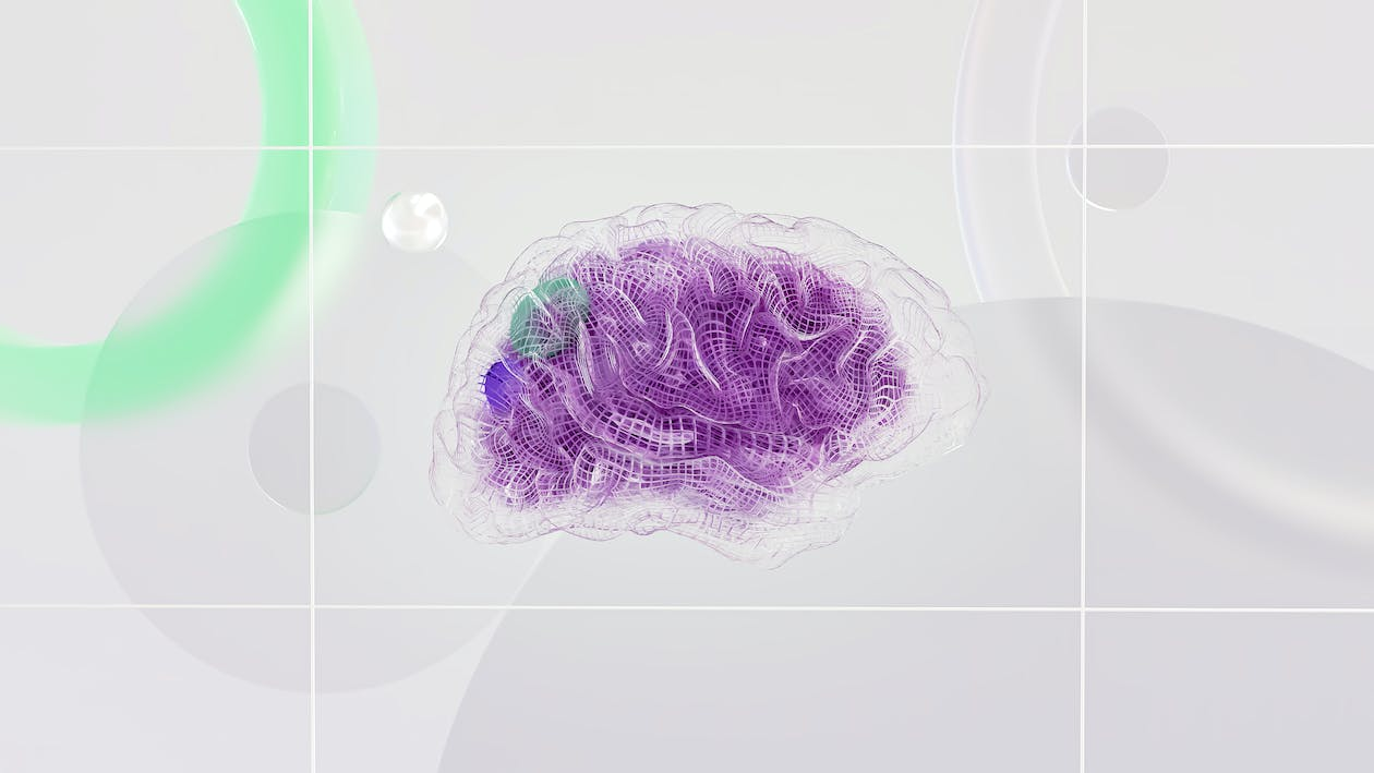
Brain mapping is a set of revolutionary techniques predicated on the mapping of properties or (biological) quantities onto spatial representations of the human brain, resulting in maps. It is a tool that neuroscientists and doctors use to evaluate brainwave neuron signals. Neurons are special cells in the brain that receive and send signals. The communication of signals can be shown in a brain map where the impulses allow a visual representation of brain activity to be created, explains Dr. Curtis Cripe.
 |
Image source: images.pexels.com |
Positron emission tomography (PET) is another common tool used for brain mapping, adds Dr. Curtis Cripe. PET is used to evaluate the metabolic function of the brain and any conditions that cause deterioration of mental function. In this type of nuclear medicine, a small dose of a radioactive substance called a radiopharmaceutical is injected through an IV. This substance enables the imaging scan to show a contrast of the tissues that are to be evaluated.
Computer axial tomography, also known as a CT or CAT scan, is a noninvasive scan that uses X-rays to create images of the brain, explains Dr. Curtis Cripe. The scan and X-ray move around the patient, allowing several images of the brain to be captured. This allows views of the brain at different angles
and depths.
 |
Image source: images.pexels.com |
QEEG is a neuro-imaging technique that is fast enough to measure neuro-function down to 100th of a millisecond, which more closely approximates brain processing speed. These faster recordings allow clearer functional measurements of brain performance with regard to thinking and processing information.
Learn more about NTL Group's research and development head Dr. Curtis Cripe and the work he does by clicking this link.